
Acne • Marks
Acne Marks: What It Is and How It Differs from Indented Scars
The acne has already settled, but a red or brown mark is still sitting in the same place months later. Acne can leave two different things behind: tissue that has been lost, and color that has stayed. They look alike in the mirror, and that is exactly why treatment splits on which one it is.

What is Acne Marks?
The acne itself has passed, but a patch of redness or a brown spot is still there. Nothing has been carved out of the skin — only the color has stayed.
The color change acne leaves behind is a separate phenomenon from the process that builds a scar. It can appear before scarring does, or alongside it. That is why marks and scars are so often mixed together on one face.
Marks then divide again. One kind is the red mark (post-inflammatory erythema, PIE). It is the pink to red discoloration left where inflammatory acne has been, and it is not pigment at all — it is the vascular change remaining in that spot showing through the skin.
The other kind is the brown mark (post-inflammatory hyperpigmentation, PIH). Inflammation drives melanin to be overproduced and scattered unevenly. Acne is not the only cause; eczema, trauma and dermatologic procedures can leave the same mark. It is more common in darker skin tones, with the literature reporting an incidence that can reach 65% in acne patients with darker skin.
Brown marks divide once more, by the layer the melanin sits in. Melanin held in the epidermis reads as brown and fades comparatively well. Where the inflammation was severe or long-running, melanin is taken up by macrophages in the dermis; that pigment reads slate-gray and lasts far longer. Because the course depends on which layer the pigment is in, the shade of the mark is one of the clues read during consultation. In the literature, Wood's lamp examination is used alongside it.
How is it different from Indented Scars?
Marks and indented scars come out of the same acne, but what they leave behind is different. A mark is color that has stayed; an indented scar is tissue that is gone. An atrophic (indented) scar is a depression in the dermis formed when collagen is lost faster than it is made, and in histopathology work on skin of color, sparse and disorganized collagen with reduced elastic tissue was seen in most of the specimens examined.
They are hard to tell apart because early on the two overlap. A freshly formed indented scar still carries redness and pigment of its own, so the depression is not easy to see until the color settles. In the other direction, a deeply colored mark can read like a shadow and be taken for a dent.
This is also where the direction of treatment parts. Treatments aimed at pigment or at blood vessels are used to lighten color, but they do not put back tissue that is gone. A depression needs treatment on the side that rebuilds tissue. Deciding what is actually left behind therefore comes first.
Indented Scars
- What is left behind
- Tissue has been lost.
- What happened in the skin
- Collagen loss outpaces synthesis and the dermis becomes depressed.
- Over time
- Usually stays as it is.
- What treatment aims at
- Rebuilding the depressed tissue.
Acne Marks
- What is left behind
- The tissue is intact; the color has stayed.
- What happened in the skin
- Pigment or vascular change left behind by inflammation remains.
- Over time
- Many fade, but it takes several months or longer.
- What treatment aims at
- The pigment or the blood vessels that remain.
The Other’s Exclusive Solution — Hybrid Injection for Acne Marks
Treatment splits on whether what remains in the mark is pigment or blood vessels.
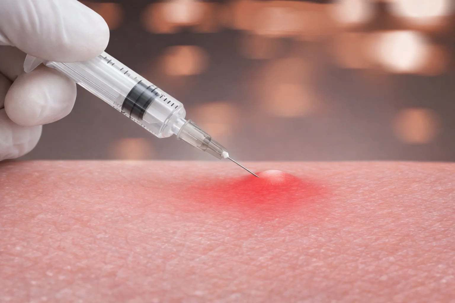
The first step in that order is settling the inflammation that caused it. The framework the literature sets out for post-inflammatory hyperpigmentation rests on three things: treating the underlying inflammatory disease, topical lightening agents, and daily broad-spectrum sunscreen. If acne is still erupting, erasing color alone simply produces a new mark next to the one just cleared, so active lesions are addressed first.
For brown marks, topical lightening agents are the center of treatment. Hydroquinone is the representative agent, and a triple combination of hydroquinone 4%, tretinoin 0.05% and fluocinolone acetonide 0.01% is commonly used. Topical retinoids and azelaic acid address acne and pigment together. Pigment-targeting lasers (Q-switched ruby, Q-switched Nd:YAG, picosecond lasers) and chemical peels are also used, but the literature attaches a caveat to all of them: they can irritate the skin and cause further pigmentation, so they call for a skilled operator and a cautious hand.
For red marks, vascular lasers are used. The principle is selective photothermolysis — a wavelength well absorbed by hemoglobin in the blood is delivered in a pulse shorter than the thermal relaxation time of the target structure, so the heat injury stays confined inside that vessel. The 585–595 nm pulsed dye laser has been used for post-inflammatory erythema, with improvement reported.
Slate-gray pigment trapped in the dermis is a different situation. Physical treatments such as peels, lasers and IPL may help pigment in the epidermis but do nothing for pigment in the dermis, and by injuring the epidermis they can make the color darker instead. Treatment is therefore not decided on color alone: which layer the pigment sits in, and whether what remains is pigment or vessels, is established first. Whatever is done, sun protection is part of the treatment. Without adequate photoprotection, recurrence is common.
01 Diagnosis — Multi-Spectrum Analysis
Analyzes whether the mark is vascular (PIE) or pigmentary (PIH), along with its depth by skin layer.
02 Prescription — Custom Mixing
Precisely mixes the ratio of inflammation-control agents and regenerative ingredients according to the severity of each patient's marks.
03 Treatment — Laser & Injection
Combines vascular and pigment lasers with The Other's dedicated injection treatment.
04 Regeneration — Post-Recovery Care
Combines hyperbaric oxygen or regenerative care to help the injected ingredients activate within the skin.
What to expect and how long it takes
How a brown mark runs depends on the layer the pigment is in. When it sits in the epidermis, the literature describes it as usually clearing or improving markedly within 6 to 12 months. When it sits in the dermis, it improves slowly and may be permanent. Even marks that resolve on their own can take months to years, and treatment often has to continue through that stretch.
Red marks also tend to fade with time, and that is where they differ from a scar that stays. How long it takes, though, varies widely from person to person depending on how severe the inflammation was and how consistently the skin was protected from light. Session counts and recovery times are not written as numbers on this page because those values change with the type of mark and the layer the pigment is in. The actual plan is set in consultation, with the lesion in front of us.
Looked at first
The color of the mark
Red marks (PIE) and brown marks (PIH) are approached differently.
Used together
Laser + injection
A laser alone does not switch off inflammation that is still running.
Matters most
Sun protection
Ultraviolet light during healing lays the pigment back down.
Aftercare
Sun protection is the foundation of caring for marks. Pigmented patches darken with sun exposure, and recurrence is common without adequate photoprotection. Daily broad-spectrum sunscreen appears in the literature as one pillar of treatment. Sunscreen does not, however, block visible light well on its own, and in skin that pigments easily, tinted formulations containing iron oxides have been reported to give better protection against visible-light-induced pigmentation.
After a vascular laser, the literature advises cooling the area immediately afterward, avoiding rubbing and gritty cleansers for several days, and staying out of the sun. Until the skin has healed, swimming, saunas, hot baths and contact sports are also avoided. Precautions matched to the treatment actually received are given afterward.
The marks that last longest are usually the ones hands have made. The extra damage of picking or squeezing active acne raises the chance that a scar forms. Treating acne appropriately and on time while it is active is understood to help reduce both how often marks and scars appear and how severe they become.
What changes by area
Facial skin and trunk skin are different to begin with. On the trunk, the thickness and structure of the stratum corneum, sebum output, the microbiome, surface pH and sweat gland distribution all differ noticeably from the face; the stratum corneum is thicker, while sebaceous gland density and activity are lower.
Acne on the back and chest adds friction and occlusion from clothing on top of that. Inflammation therefore runs deeper, scarring risk is higher, and penetration of topical medication is limited, so an approach that worked on the face does not always carry over. These differences are reported in the literature for the course of acne itself and for the response to topical treatment. Marks are what that acne leaves behind, so how they settle can differ by site as well.
Other conditions that look similar
Some things look like marks but are a different pigmentary disorder. The differential diagnosis for post-inflammatory hyperpigmentation includes melasma, solar lentigines, tinea versicolor, acanthosis nigricans, lichen planus pigmentosus, macular amyloidosis, ochronosis, erythema dyschromicum perstans and discoid lupus erythematosus. Pigment whose distribution does not match where the acne was is a reason to look at other causes as well.
The same holds for redness. Post-inflammatory erythema stays exactly where the inflammation was. If redness on the face does not follow the distribution the acne took, causes other than marks are checked alongside it.
Frequently asked questions
How do I tell an acne mark from a scar?
It is hard to separate them by appearance alone. Early on, a fresh scar still carries redness and pigment, so the depression is not obvious, and a deeply colored mark can read like a shadow and be mistaken for a dent. The real dividing line is whether a contour remains after the color has faded, and that is confirmed in consultation.
Will it go away if I leave it alone?
Color changes left by acne often improve with time, but it takes several months or longer. Brown marks sitting in the epidermis usually clear or improve markedly within 6 to 12 months in the literature, while slate-gray pigment trapped in the dermis improves slowly and may be permanent. A scar where tissue has been lost, by contrast, does not resolve on its own.
Why are red marks and brown marks treated differently?
Because what remains is different. A red mark is a vascular change; a brown mark is melanin. Vascular lasers use wavelengths absorbed by hemoglobin, while pigment treatment targets melanin. Both kinds of mark often sit on the same face, so the order of treatment follows whichever is the dominant problem.
Do I really have to wear sunscreen?
Yes. Daily broad-spectrum sunscreen is listed in the literature as one of the pillars of treating post-inflammatory hyperpigmentation. Pigmented patches darken with sun exposure, and recurrence is common without adequate photoprotection. In skin that pigments easily, tinted formulations that also screen visible light have been reported to give better protection.
Can a procedure itself create more marks?
It can. Lasers, light-based treatments and chemical peels are themselves listed in the literature as causes of post-inflammatory hyperpigmentation. That is why skin tone, the layer the pigment sits in, recent sun exposure and current medications are checked before the intensity and the interval are set.
Why is a slate-gray mark so hard to clear?
Because the melanin is trapped inside dermal macrophages rather than in the epidermis. Peels, lasers and IPL that target epidermal pigment have no effect on dermal pigment, and by damaging the epidermis they can make the color darker. The literature regards improvement in these cases as slow and possibly permanent.
This page is general information about a treatment and is not a diagnosis or a recommendation. The right choice differs by skin condition, so treatment is decided after an in-person examination. Individual results vary.
References
- Acne scarring DermNet NZ
- A cross-sectional pilot study evaluating the histopathology of atrophic acne scars IJDVL
- Easy as PIE (postinflammatory erythema) J Clin Aesthet Dermatol 2013;6(9)
- Post-inflammatory hyperpigmentation StatPearls
- Pulsed dye laser treatment DermNet NZ
- Photoprotection beyond ultraviolet radiation: a review of tinted sunscreens PubMed
Related information
Treatment pages
- Raised (papular) acne scars — When what is left is a raised scar rather than a mark
- The difference between acne marks and indented scars — How marks and scars are told apart
- Treatment for indented acne scars — What is done when tissue has been lost
- Facial redness and rosacea flushing — When the redness does not match where the acne was