
Scar • Keloid
Keloid: What It Is and How It Differs from Hypertrophic Scar
A scar on the chest, the shoulder or the earlobe sometimes grows larger than the wound that caused it, and it itches or hurts. A scar that behaves this way is called a keloid. Unlike a hypertrophic scar, which stays inside the borders of the original wound, a keloid spreads into the normal skin around it and does not settle down on its own. This page explains what separates the two, and why a keloid is a condition in which recurrence has to be managed alongside the scar itself.
What is Keloid?
A spot where acne once sat, a piercing hole in the ear, a surgical line — instead of healing closed, it grows larger and firmer as the months pass. It may itch, and it may hurt. Scar tissue that keeps growing instead of resolving is called a keloid.
What decides whether a raised scar is a keloid is not its size and not its colour but the border. If the tissue grows past the edge of the original wound into surrounding normal skin, it is a keloid; if it has thickened but stayed within that edge, it is by definition not one. The literature treats this horizontal growth as the central basis for the diagnosis.
A keloid starts where skin was injured. Surgery, burns, insect bites, vaccinations and piercings are typical triggers, and so are acne and folliculitis. A hypertrophic scar usually appears within a few weeks of the injury; a keloid can rise months or even years afterwards.
It does not occur with equal frequency in everyone, and that is part of the picture. Prevalence is highest in people of African descent, followed by Asian and Hispanic populations, and is lowest in white populations. Incidence rises with darker skin (Fitzpatrick III–VI), and self-reported figures of 16% have been reported in Black populations. It appears most often between the ages of 11 and 30, and reports of keloids in twins and across several generations of the same family point to a genetic predisposition. Prevalence figures specific to Korean patients, however, are something we were not able to source.
The most common accompanying symptom is itch, followed by pain. Keloids are common on the earlobe, shoulder, chest, back, cheek and knee; they are uncommon on the face, though they can appear along the jawline.
How is it different from Hypertrophic Scar?
Both are states in which too much scar tissue has been laid down, so on the surface both look similarly red, firm and raised. It is often hard to tell them apart from the outside, and there are cases where a photograph alone is not enough either. What separates them is the direction they grow and where they end up over time.
A hypertrophic scar stays within the injured area and can regress as time passes. A keloid crosses that border into normal skin and does not regress on its own. This horizontal growth is the strongest reason the literature has kept the two classified separately.
They also differ inside the tissue. Collagen in a hypertrophic scar is arranged in relatively regular wavy bundles, while collagen in a keloid shows no clear pattern; collagen production is reported at roughly 3 times normal skin in hypertrophic scars and roughly 20 times normal in keloids. On biopsy, thick hyalinised keloidal collagen bundles are seen on the keloid side. The distinction matters because the outlook differs — with one there is room to wait and watch, with the other recurrence has to be managed even after treatment.
Hypertrophic Scar
- Wound border
- Stays within the injured area
- Natural course
- Can regress over time
- When it appears
- Within a few weeks of the injury
- Collagen
- Regular wavy arrangement · about 3 times normal
- Predisposition
- No clear association with ethnicity
Keloid
- Wound border
- Crosses the border into normal skin
- Natural course
- Does not regress on its own
- When it appears
- Months to years after the injury
- Collagen
- No clear arrangement pattern · about 20 times normal
- Predisposition
- Higher incidence in darker skin
How is it treated?
A keloid is not a lump sitting on top of the skin but chronic inflammation held in the reticular dermis.
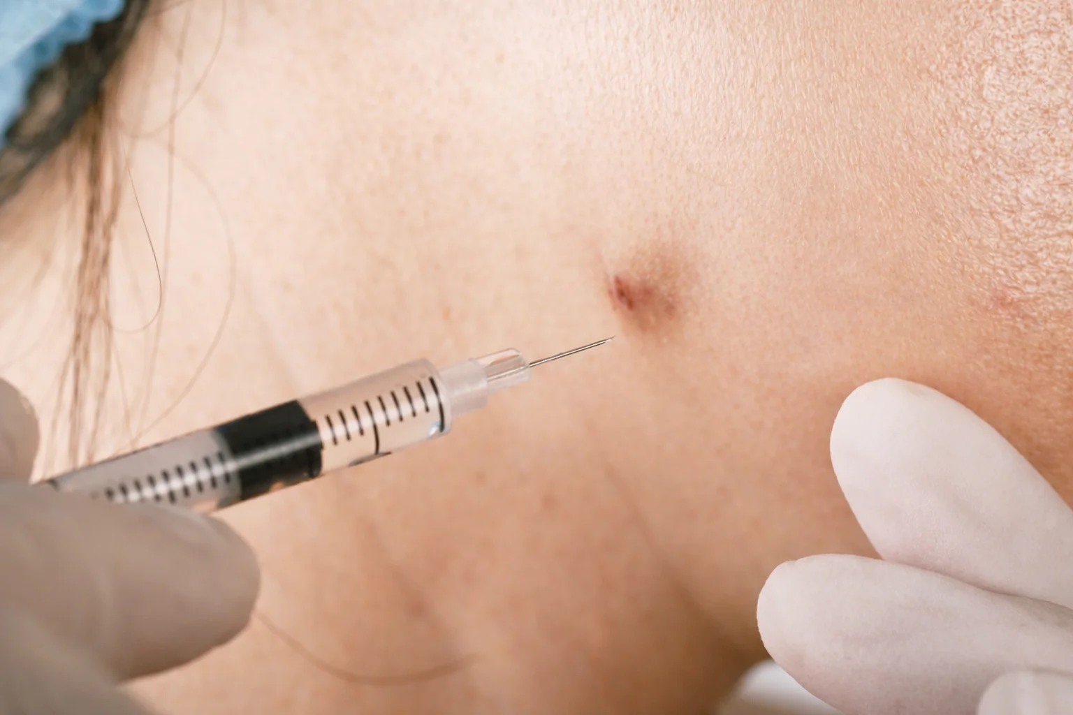
Normal wound healing runs through an inflammatory phase, a proliferative phase and a remodelling phase, and then it stops. In a keloid that control is lost: extracellular matrix proteins such as collagen, elastin and proteoglycans are overproduced, and the numbers of fibroblasts and mast cells rise. Overexpression of TGF-β1 and TGF-β2 together with reduced expression of TGF-β3 is thought to drive the increase in extracellular matrix.
This is also why keloids do not form just anywhere. The reticular dermis is where inflammatory cells, fibroblasts, newly formed vessels and collagen deposition gather, and when repeated skin tension is added on top, the inflammation worsens and lasts longer. So keloids clearly favour the anterior chest and the scapular region, where skin is constantly pulled, and are rare on the scalp vertex, the shin and the upper eyelid, where the skin has little occasion to stretch or contract.
What this clinic uses for keloids is intralesional triamcinolone (TA) injection, 5-FU injection and lasers. Triamcinolone is the intralesional steroid most widely used for keloids and is regarded as first-line; it is reported to reduce both dermal fibroblast proliferation and collagen production. In laboratory studies it suppressed cell proliferation, arrested the cell cycle and decreased type I collagen synthesis in keloid fibroblasts. In other words, it does not shave away tissue that has already accumulated — it holds down the process that keeps adding to it. What makes The Other distinctive is the 5-FU injection. Combining 5-FU brings the following advantages.
1. The effect itself is better
In several randomised controlled trials and meta-analyses, the combination group was superior in reduction of lesion height, in pliability, in redness and in VSS/POSAS score improvement. The response also comes sooner, so relief of symptoms such as itch and pain arrives earlier.
2. The recurrence rate is lower
At 6 to 12 months of follow-up, the recurrence rate in the combination group is reported to be significantly lower. With TA alone the early response can be good but recurrence is the problem, and because 5-FU suppresses fibroblast proliferation itself, that is where the difference appears.
3. Steroid side effects are reduced
This is the part felt most in practice. Mixing in 5-FU lowers the cumulative TA dose, so skin atrophy, telangiectasia, hypopigmentation and satellite lesions around the margin decrease markedly. It matters especially in large lesions that need repeated injection.
4. The mechanisms are complementary
TA works through anti-inflammatory action and through activation of collagenase by inhibiting α2-macroglobulin, while 5-FU, as a pyrimidine analogue, directly suppresses proliferating fibroblasts and blocks TGF-β-induced COL1A2 expression by inhibiting Smad3 phosphorylation. It is therefore particularly useful in steroid-resistant lesions and in recurrent keloids.
Lasers have been used alongside injection, targeting the redness, the height and the pliability of the scar. Which wavelength is used and under what settings changes both what is being targeted and what risk has to be accepted along with it. Which device is combined here, and for what purpose, is decided in an in-person examination after looking at the thickness and site of the lesion and the patient's skin colour.
What to expect and how long it takes
A keloid is less a condition that a single procedure finishes off, and more one in which the size and the symptoms are brought down and recurrence is managed.
In the literature, intralesional triamcinolone is given alone or as an adjunct at intervals of 4 to 6 weeks over several months (some sources give 4 to 8 weeks). The range of treatment response differs greatly between studies, and recurrence after injection is reported at 9 to 50%. The literature itself notes that randomised controlled trials of intralesional steroids are incompletely reported and highly heterogeneous, so the course cannot be promised as a single number.
Surgical excision is in a similar position. Recurrence after excision alone is reported at 50 to 80%, and adjuncts such as steroid injection or radiation are known to be able to reduce it.
The actual injection interval, the total number of sessions and the point at which change becomes visible depend on the thickness and site of the lesion and on how previous treatment went, so they are not written here as a single figure. They are set in an in-person examination.
Aftercare
What happens between injections often decides the course. The following are general methods cited in the literature and in international recommendations; which of them applies, and how, is decided in consultation according to the lesion and the site.
Silicone sheets and silicone gel are what international scar-management recommendations list as first choice for both prevention and treatment of hypertrophic scars and keloids. The recommended regimen is to apply them after the wound has fully closed, for at least 12 hours a day over 2 to 3 months; some sources give 12 to 24 hours a day for 8 to 12 weeks or longer.
Pressure therapy requires 23 hours of wear a day and is most effective immediately after injury or surgery. Silicone combined with pressure has been reported to give better improvement than either alone, while a meta-analysis found the evidence for preventing abnormal scarring in high-risk groups to be weak. Individual results vary.
Detailed care after a procedure — washing, sun exposure and topical ointments — follows the instructions given at the clinic.
What changes by area
Keloids are selective about where they form. They clearly favour the anterior chest and the scapular region, where skin tension is constant and repeated, and they take on shapes characteristic of the site — a butterfly shape on the shoulder, a crab-claw shape on the anterior chest, a dumbbell shape on the upper arm.
Conversely they almost never form on the scalp vertex or the shin, where skin rarely stretches or contracts, and the upper eyelid — which stays relaxed whether the eye is open or closed — is another uncommon site. The earlobe is a common site, often after a piercing. The jawline is uncommon for the face, but a keloid can form where acne and folliculitis kept recurring.
Other conditions that look similar
Not every raised scar is a keloid. A hypertrophic scar stays within the wound border and can regress over time, so it is approached differently. Papular scars — the white or skin-coloured bumps that rise around the nose and chin after acne — are neither red nor itchy and change little in size, and they are approached differently again from keloids.
Red marks and brown pigment left behind where acne has passed are flat changes that are not raised at all; those are marks rather than scars. Because what needs to be done is the opposite depending on which one it is, the starting point is always telling them apart.
Frequently asked questions
Will a keloid go away on its own?
Keloids are reported not to regress on their own. A hypertrophic scar, which stays inside the wound border, can regress over time; a keloid, which grows past that border, does not. There are cases where the area widens while the patient waits for it to improve.
How is it told apart from a hypertrophic scar?
The criterion is not size or colour but the border. If the tissue spreads past the edge of the original wound into normal skin, it is regarded as a keloid; if it has thickened only within the wound site, it is closer to a hypertrophic scar. This horizontal growth is what the literature uses as the central basis for diagnosing a keloid.
Are keloids hereditary?
Occurrence in twins and across several generations of the same family has been reported, which points to a genetic predisposition. Incidence is higher in darker skin, and keloids appear most often between the ages of 11 and 30. That said, we were not able to confirm a single test that decides whether someone is predisposed, well enough to write it on this page. The judgement is made together with family history and how previous scars behaved.
Can acne cause a keloid?
Acne and folliculitis are named in the literature as triggers for keloids. Keloids following acne or folliculitis are especially common on the anterior chest, where skin tension is applied repeatedly. But not every raised lump left at an acne site is a keloid, so telling them apart comes first.
Can it simply be cut out?
Recurrence after excision alone is reported at 50 to 80%. Adjuncts such as steroid injection or radiation are known to be able to reduce recurrence. Whether to excise, and which adjunct to combine, is decided together with the size and site of the lesion and how previous treatment went.
Will repeated injections thin my skin?
Skin atrophy, telangiectasia and pigment change are known risks of intralesional steroid. The size of the risk depends on the concentration and dose of the steroid and on the depth of injection, and one survey reported atrophy persisting beyond 6 months. That is why 5-FU is combined, so that treatment keeps the total steroid dose to a minimum.
This page is general information about a treatment and is not a diagnosis or a recommendation. The right choice differs by skin condition, so treatment is decided after an in-person examination. Individual results vary.
References
- Hypertrophic Scarring Keloids StatPearls
- Hypertrophic scars and keloids: Overview of the evidence and practical guide for differentiating between these abnormal scars Exp Dermatol 2021
- Hypertrophic scars and keloids DermNet NZ
- Keloid and hypertrophic scars are the result of chronic inflammation in the reticular dermis Int J Mol Sci 2017
- A prospective study of patients presenting with pre-sternal keloids NCBI PMC
- Laser treatment of specific scar characteristics in hypertrophic scars and keloid: A systematic review JPRAS 2020
- Fractional carbon dioxide, long pulse Nd:YAG and pulsed dye laser in the management of keloids J Cutan Aesthet Surg
- Intralesional steroid injection DermNet NZ
- Intralesional corticosteroid administration in the treatment of keloids: a scoping review on injection method Dermatology (Karger)
- An evaluation of evidence regarding application of silicone gel sheeting J Clin Aesthet Dermatol
- KENALOG-40 product label DailyMed
Related information
Treatment pages
- See the papular scar treatment page — Care for the white bumps that rise on the nose and chin after acne
- See the acne marks treatment page — A different trace left behind by the same acne
- Telling acne marks and acne scars apart — Flat marks and raised or indented scars are approached differently
- See all acne and scar treatments — Treatment guidance by type of scar