
Special Clinic • Milia
Milia: What It Is and How It Differs from Syringoma
- Written by
- The Other Dermatology medical team
- Published
- 2026.08.21
Small white grains settle under the eyes, on the cheek or beside the nose, and they feel firm and distinct under the fingertip. They look as though a squeeze would pop them out, but the same areas also grow other lesions that overlap with them in appearance — syringoma and sebaceous hyperplasia among them. This page sets out what milia are, why the course described for newborns differs from the one described for adults, and how far the confirmed evidence goes before it stops.
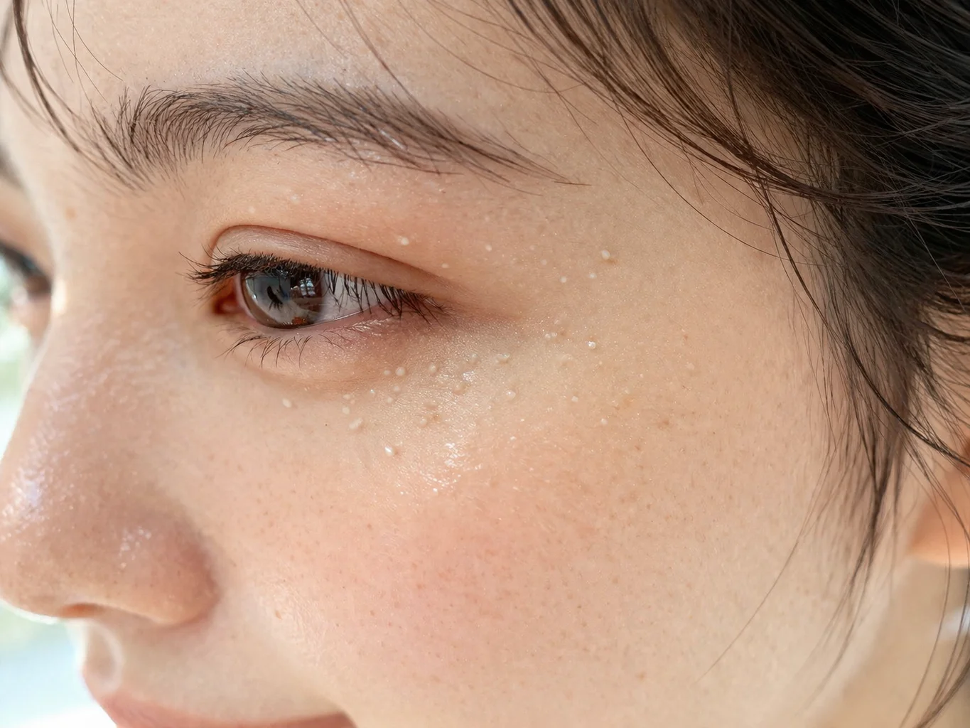
What is Milia?
What you see in the mirror is a white, millet-like grain under the eye, on the cheek or beside the nose. It is not red the way acne is, it catches under the fingertip as a firm grain, and its shape does not change much from one day to the next.
In medical terms, milia (singular: milium) are benign cysts filled with keratin that sit beneath the epidermis. The literature records them as subepidermal keratin cysts and classifies them as pseudocysts formed because keratin cannot get out. They are smooth and dome-shaped, white to pale yellow, and on darker skin they can carry a faint bluish cast.
Reported size differs by source. StatPearls describes congenital milia as under 3 mm, while DermNet records milia as superficial papules of 1–2 mm. Either way these are small lesions of a few millimeters or less, but this is not a figure that can be collapsed into one number.
Milia are divided into primary and secondary. Most primary milia are congenital, present from birth, and appear on the nose, scalp, eyelids and cheeks, along the gum border (Bohn nodules) and on the palate (Epstein pearls); acquired milia that arise later without a particular cause favor the eyelids, cheeks, forehead and genitalia. Primary milia are thought to arise from the sebaceous portion of the vellus hair follicle.
Secondary milia follow something that happened at the site. The literature lists trauma such as dermabrasion and radiotherapy, prolonged topical corticosteroid use with underlying atrophy, use of non-steroidal anti-inflammatory drugs, and the aftermath of bullous disorders once an inflammatory skin disease has settled. They arise where the epidermis is repairing itself after blistering or injury, and epidermolysis bullosa, bullous pemphigoid, porphyria cutanea tarda, thermal burns and the period after dermabrasion are named together.
Histologically, milia are classified as benign lesions. The literature treats them as benign and asymptomatic and records no reported systemic complications. That statement, however, is about lesions confirmed to be milia. Not every small white bump under the eye is a milium, so what it is gets decided at an examination.
How is it different from Syringoma?
The two lesions turn up in the same places. Milia favor the eyelids, cheeks and forehead, and syringomas cluster around the eyes, especially on the lower lids. Size does not separate them either — milia are recorded as under 3 mm (StatPearls, describing congenital lesions) or 1–2 mm (DermNet), and syringomas as 2–4 mm (StatPearls), so the ranges overlap. We could not find a source that measured adult milia separately. Size alone does not tell the two apart.
What separates them is what is sitting in that spot. A milium is a cyst formed around trapped keratin, so there are contents to empty; a syringoma is a benign tumor of proliferating sweat ducts, so there is tissue to remove. The target of removal differs. This is not a contrast that resolves into depth — the literature records that the proliferation in syringoma is restricted to the superficial dermis. Milia sit beneath the epidermis and syringomas in the superficial dermis just below it, which is not a relationship that can be sorted into a shallow one and a deep one.
It is also worth noting that the lesion the literature names first in the differential for milia is not syringoma but sebaceous hyperplasia, with comedonal acne, flat warts and milia-like idiopathic calcinosis cutis alongside it on the list. In the other direction, milia do appear on the differential list for syringoma. This page places syringoma beside milia because the two are seen together in the same area, which is another way of saying that a small bump under the eye is hard to judge by appearance. What it is gets decided at an in-person examination.
Syringoma
- What it is
- A benign tumor of proliferating sweat ducts
- Origin
- Arises from the eccrine sweat ducts
- Where it sits
- Superficial dermis
- Target of removal
- The proliferated sweat-duct structure
- Usual location
- Around the eyes, especially the lower lids
Milia
- What it is
- A keratin-filled cyst beneath the epidermis
- Origin
- The sebaceous portion of the vellus hair follicle (primary milia)
- Where it sits
- Beneath the epidermis
- Target of removal
- The keratin cyst itself
- Usual location
- Eyelids, cheeks, forehead
How is it treated?
Milia are handled by emptying the trapped keratin, not by destroying tissue.
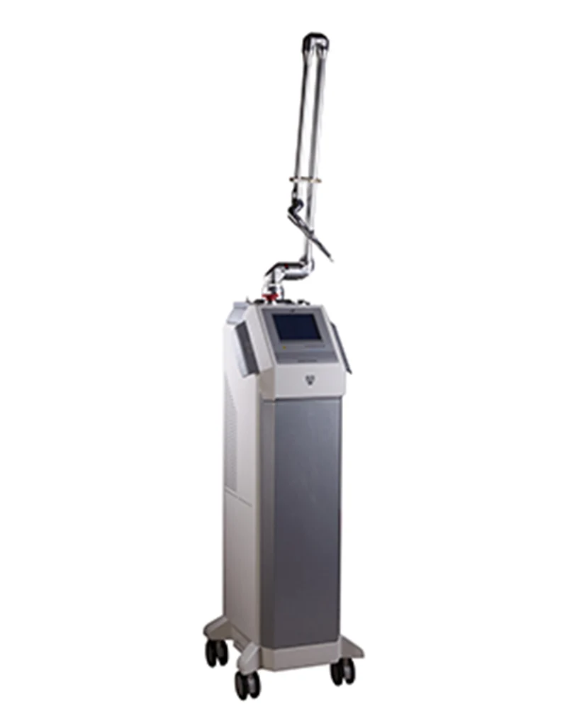
The removal methods the literature lists are simple in-office procedures that evacuate the contents with an instrument, with topical retinoids and electrodesiccation or electrocautery named alongside as options when there are many lesions. The specific steps are not reproduced on this page — they are not the sort of thing to set down in a form that can be read and copied at home. We could not find a source stating which retinoid, at what concentration, for how long, so that is not written here either.
This clinic’s own treatment guide states that “a very tiny opening is made at the entrance of the keratin cyst using a CO2 or Er:YAG laser” and that “only the contents are cleanly removed without applying excessive force to the surrounding tissue”. How many visits it takes is not given as a figure on this page — the literature holds no number that can stand as a usual session count, and the figure for this clinic’s procedure could not be confirmed either.
Laser does not appear on the literature’s list of removal methods. What could be confirmed about laser is a single case report in which CO2 laser vaporization was performed on 4 patients (aged 12–50) with several variants of milia and followed for 12–36 months; the authors write that the patients showed "marked improvement" after a few sessions. The design had no control group and no quantitative rating scale, and the subjects were rare variants rather than ordinary adult milia.
No randomized controlled trial or systematic review of milia treatment could be found. The authors of that case report state themselves that an optimal treatment modality has not been established. The literature also records that treatment is not required unless the milia are a concern to the patient — what this page describes is not a procedure that has to be done.
What to expect and how long it takes
The literature describes milia in newborns and milia in adults differently. Congenital milia are common enough to be read as a normal variant, appearing in 40–50% of healthy full-term newborns, and they are recorded as clearing on their own without scarring within a few weeks to a few months, usually during the first month of life. In preterm infants they may appear later.
For acquired milia in adults, the literature records that they may persist unless treated. The course seen in newborns, where the lesions resolve by themselves, does not transfer directly to adults. No source measuring what proportion resolve spontaneously in adults could be found.
On whether a removed milium returns to the same spot, no evidence confirmed well enough to put on this page could be found. What has been reported are individual cases of one to four patients, and their conclusions disagree with one another. A removed lesion returning and a new lesion appearing elsewhere are also two different questions, and the literature gives a figure for neither.
Time to visible change, how long it lasts, a usual number of sessions and a recovery period are not given as figures on this page. The literature holds no number that can stand as a usual session count, and the figure for this clinic’s procedure could not be confirmed either. It varies with the number of lesions, the site and the state of the skin, and is settled at an in-person consultation. Individual variation applies.
The risks that come with the procedure belong here beside the course. Infection is reported even in professional procedures performed with instruments — in a report covering 65 patients who underwent facial dermabrasion, secondary infection occurred in 2, and in a retrospective analysis of 25 patients who had laser resurfacing for actinic keratoses, infection was reported at 12%. Scarring appears in that same dermabrasion report as small deep scars in 8 patients, and in the resurfacing analysis as scar formation at 8%. Around the eyes the weight of that is different — one case report records that in this area even minimal scarring can result in ocular complications. A small lesion does not make the procedure a light one. For pigment change, both hypopigmentation and hyperpigmentation are reported, but the rates are not carried onto this page — every citable figure comes from procedures that resurface the whole face, measured in a population different from the one this page is about. Where the skin is darker, the literature recommends discussing the possibility of pigment change in advance. All of the figures above measured procedures that treat the entire face rather than milia removal, but the fact that infection and scarring do not disappear even with sterile instruments in a clinical setting stands as it is.
The literature records that no systemic complications have been reported for milia themselves. That is a statement about the lesion, and it does not mean that the risk of the procedure removing it is small. No evidence on the safety of the procedure during pregnancy or breastfeeding could be found — with no grounds to write here that it is either safe or unsafe, the timing is settled in consultation.
What changes by area
Milia arising spontaneously in adults favor the eyelids, cheeks, forehead and genitalia, the literature records. Of those, the area around the eyes is also where syringomas cluster on the lower lids, so the two lesions share the site and share the size range as well. Under the eye, appearance alone does not settle what a lesion is.
The eye area is a careful site in its own right. One case report (1 patient) records that in this region even minimal scarring can result in ocular complications. It is not a source that carries great evidential weight, but neither is it a statement that gives any reason to rate the risk lower.
In newborns, milia appear on the nose, scalp, eyelids and cheeks, and are also seen along the gum border (Bohn nodules) or on the palate (Epstein pearls). That is the setting the literature describes as requiring no specific treatment.
Other conditions that look similar
Not every small white bump under the eye is a milium. The lesion the literature names as the principal differential for milia is sebaceous hyperplasia, which presents as white-to-yellow papules grouped around the upper lip and nose. Comedonal acne, flat warts and milia-like idiopathic calcinosis cutis are also on the differential list.
Lesions that are new after a procedure are looked at alongside secondary milia. In a study of 100 patients who had fully ablative or fractional CO2 laser resurfacing for cosmetic purposes, milia were observed in 5.1% of the group; in a retrospective analysis of 25 patients who had resurfacing to treat actinic keratoses, the figure was 12%; and among 65 patients who underwent facial dermabrasion, milia were reported in 20. The three values measured different procedures in different populations, so they cannot be pooled or averaged, and none of them measured a procedure that removes milia.
Milia are also reported after burns, skin grafting and prolonged topical corticosteroid use, and at sites where a bullous disorder such as epidermolysis bullosa, bullous pemphigoid or porphyria cutanea tarda has passed through. If lesions have appeared recently and are concentrated in one area, what happened at that site is looked at along with them.
Frequently asked questions
Are milia a dangerous condition?
The literature treats milia as benign, asymptomatic lesions and records no reported systemic complications. That statement, though, is about lesions confirmed to be milia. Not every small white bump under the eye is a milium, so what it is gets decided at an examination.
Do they have to be treated?
The literature records that milia need no treatment unless they are a concern to the patient. They are not lesions that cause pain or loss of function, so watching them is an available choice. Congenital milia are likewise recorded as requiring no specific treatment. That said, if lesions persist for a long time or are widely spread, another cause may need to be checked before removal — particularly when other skin findings are present, and in that case repeatedly clearing the visible lesions may not be the answer. Where the cause is identifiable and secondary — trauma, burns, dermabrasion or radiotherapy, prolonged topical corticosteroid use, or the site of a past bullous disorder — lesions can return to the same place unless that cause is addressed as well.
Do the white grains on a baby’s face go away on their own? Is it the same for adults?
Congenital milia are common enough to appear in 40–50% of healthy full-term newborns, and they are recorded as clearing within a few weeks to a few months without scarring, usually during the first month of life. Adults are different. The literature records that acquired milia may persist unless treated. The course seen in newborns does not transfer directly to adults.
Can I squeeze them at home?
The steps are not written on this page. No source measuring the risk of squeezing milia yourself could be found, but the general guidance from the professional body is that squeezing a skin lesion pushes the contents further inward, worsens inflammation, and carries a risk of infection from bacteria on the hands and of scarring (the condition that guidance addresses is acne). For periocular milia there is also a case report (1 patient, a rare variant) recording that manual expression is often followed by local recurrence. On top of that, a bump under the eye overlaps in appearance with syringoma and sebaceous hyperplasia, so you would be working on something without knowing what it is. What it is gets decided at an in-person examination.
Will they come back after removal?
No evidence confirmed well enough to answer this with a number could be found. No study calculating a recurrence rate for milia was identified in the medical databases; what has been reported are individual cases of one to four patients, and their conclusions disagree. A removed lesion returning to the same spot and a new one appearing elsewhere are also two different questions.
How do I tell whether a bump under my eye is a milium or a syringoma?
Appearance alone does not separate them. The two overlap in location around the lower eyelid and overlap in size range as well. What differs is what is sitting in that spot: a milium is a cyst of trapped keratin, and a syringoma is a benign tumor of proliferating sweat ducts. That distinction is settled by examination.
This page is general information about a treatment and is not a diagnosis or a recommendation. The right choice differs by skin condition, so treatment is decided after an in-person examination. Individual results vary.
References
- Gallardo Avila PP, Mendez MD. Milia. StatPearls (last revised 2023-01-31) NBK560481 · accessed 2026-08-18
- Geist R, Crane JS. Syringoma. StatPearls (last revised 2024-03-20) NBK603740 · accessed 2026-08-18
- Gupta M, Oakley A. Milium, milia. DermNet (2009) accessed 2026-08-18
- Oakley A. Cutaneous cysts and pseudocysts. DermNet (written 1997 / updated 2016-02) accessed 2026-08-18
- Pozo Jd, Castiñeiras I, Fernández-Jorge B. Variants of milia successfully treated with CO(2) laser vaporization. J Cosmet Laser Ther. 2010 Aug;12(4):191-4 PMID 20590368 · case report, 4 patients
- Voth H, Reinhard G. Periocular milia en plaque successfully treated by erbium:YAG laser ablation. J Cosmet Laser Ther. 2011 Feb;13(1):35-7 PMID 21250787 · case report, 1 patient
- Duplechain JK, Rubin MG, Kim K. Novel post-treatment care after ablative and fractional CO2 laser resurfacing. J Cosmet Laser Ther. 2014 Apr;16(2):77-82 PMID 24131071 · 100 patients
- Ostertag JU, Quaedvlieg PJ, Neumann MH, Krekels GA. Recurrence rates and long-term follow-up after laser resurfacing as a treatment for widespread actinic keratoses on the face and scalp. Dermatol Surg. 2006 Feb;32(2):261-7 PMID 16442048 · retrospective analysis, 25 patients
- Savant SS. Facial dermabrasion in acne scars and genodermatoses — A study of 65 patients. Indian J Dermatol Venereol Leprol. 2000;66(2):79-84 PMID 20877033 · 65 patients
- American Academy of Dermatology. Pimple popping: Why only a dermatologist should do it (last updated 2023-10-17) the condition addressed is acne · accessed 2026-08-18
Related information
- Syringoma & Milia — Special Clinic — Where both lesions are differentiated and handled in one visit
- Under-eye and eye-area treatment guide — The area where milia and syringoma appear together