
Special Clinic • Syringoma
Syringoma: What It Is and How It Differs from Milia
- Written by
- The Other Dermatology medical team
- Published
- 2026.08.21
Several small, firm, skin-colored bumps sit clustered under the eye — and sometimes they have already been lasered once and come back in the same spot. Not every small bump around the eye is the same thing, though: **rarely**, malignant adnexal tumors that look benign have been reported on the eyelid, so **establishing what it is by examination comes first**. This page explains how syringoma differs from milia, and why syringoma is a condition that has to be discussed together with recurrence.

What is Syringoma?
In the mirror you see small bumps close to your own skin color under the eye — a few of them, sometimes several dozen, grouped together. They feel firm, and squeezing produces nothing. One of the lesions that looks like this is syringoma.
Syringoma is a benign adnexal tumor that arises from the ducts carrying sweat. Under the microscope, small ducts and epithelial strands sit within the dermis, and shapes described as resembling tadpoles or commas are characteristic. That classic appearance is not always present — in a review of 61 Korean patients with histologically confirmed syringoma it was seen in 34 cases (55.7%). Standard references disagree about where exactly it originates: some place it in the intraepidermal portion of the sweat duct, others describe a proliferation of sweat glands within the dermis. This page therefore goes no further than 'it arises from the ducts that carry sweat'.
Why it develops is not known. Hormonal imbalance is the leading hypothesis, but the word the literature uses is conjecture, and rare familial cases inherited in an autosomal dominant pattern have been reported, so a genetic predisposition is discussed alongside it. Although more than 800 cases have been reported over 50 years, a systematic review states that the pathophysiology remains poorly understood and treatment remains a challenge.
It is more common in women, usually appears after puberty in early adulthood, and is described as more common in people of Asian descent. Reported sex ratios differ so widely between sources that no figure is given here. The favored location is around the eyes, with a marked preference for the lower eyelid, and it also appears on the forehead, cheeks, axillae, and genital area. Typically it presents as multiple papules of 2–4 mm; in the same cohort of 61 Korean patients, the eyelid was the most common site at 43 cases (70.5%), and skin-colored was the most common color at 30 cases (49.2%). The denominator of those proportions is the 61 patients who attended that hospital's dermatology department, not a population.
It is usually asymptomatic, although it can itch. No case of a lesion confirmed as syringoma undergoing malignant change has been reported. That, however, is the story after it has been established what the lesion is; what it is confused with before that point is covered in the next section. The common form — multiple facial lesions — tends to persist unless treated, while in the eruptive variant that spreads widely over the trunk, the possibility of spontaneous regression over several years has been reported.
How is it different from Milia?
Syringoma and milia both appear around the eyes as bumps the size of a grain of millet, which is why they are often dealt with in the same consultation. The two lesions, however, are built differently. Milia are small cysts filled with keratin — a sac with contents inside. Syringoma is proliferated tissue derived from sweat ducts, that is, a tumor.
That difference decides what removal has to target. Milia are keratin trapped in a sac, so there are contents to be emptied out; syringoma has nothing to be emptied, so the proliferated tissue itself has to be destroyed, and whatever is left comes back up. Standard references state that because the lesion lies within the dermis, it can recur after superficial treatment. Covering milia in detail is not this page's job, so an article on milia is linked below. It is common to hear the two lesions separated into deep versus superficial, but that is not the axis that divides them — the proliferation in syringoma is also described as confined to the superficial dermis, and milia on the eyelids and cheeks are likewise superficial lesions.
There is also a reason they are hard to tell apart by appearance. A milium-like variant of syringoma has been described since 1987, and histologically a large keratin cyst near the epidermis is continuous with syringomatous epithelial strands below it. One literature review notes that this variant occurs more often in Asian patients, but it accompanies a single case report and is not a figure to read as a frequency.
And not every small bump around the eye is benign. Microcystic adnexal carcinoma is a rare malignant sweat gland tumor, common on the face and neck with a preference for the midface, and it can mimic syringoma histologically. What separates it from syringoma is invasion into the deep dermis and subcutis and perineural involvement — and those lie where a superficial shave biopsy does not reach, which is why a deep biopsy is recommended when this tumor is suspected. On the eyelid, three cases of syringomatous carcinoma that appeared benign have been reported; one patient required five excisions to achieve complete removal, and another recurred. This is rare, and the rarity is recorded here too. One of those cases is described as difficult to diagnose because a superficial shave biopsy may show only benign features. A procedure that destroys a lesion also destroys the opportunity to identify it — self-extraction, or removal without confirmation, carries not only the risk of scarring and infection but the risk of erasing the diagnosis. Examination comes first, and a biopsy follows if it is judged necessary. The conclusion here is not 'every syringoma needs a biopsy' but 'nothing is removed before it is established what it is' — what the literature calls mandatory is a deep biopsy when malignancy is suspected.
Milia
- What it is
- A cyst filled with keratin — a sac with contents inside
- Where it comes from
- Arises from vellus hair follicles
- Appearance
- Pearly white, just beneath the skin surface
- What removal targets
- The keratin cyst itself — there are contents to empty out
- Course after removal
- No confirmed source on the course after removal was found
Syringoma
- What it is
- A benign tumor derived from sweat ducts — proliferated tissue
- Where it comes from
- Arises from eccrine sweat ducts
- Appearance
- Skin-colored is the most common (49.2% of 61 Korean patients) · firm papule
- What removal targets
- The proliferated tissue itself has to be destroyed
- Course after removal
- Recurrence is reported regardless of the method used
How is it treated?
What has to be removed in syringoma is not contents to be emptied but proliferated tissue.
So an approach that only reaches the surface can leave the lesion behind. Standard references state that because the lesion lies within the dermis, it can recur after superficial treatment. Few studies have actually measured lesion depth; in a 1998 Korean study that analyzed 8 of the tissue specimens obtained from 45 patients, mean tumor depth was 0.70 mm (standard deviation 0.20, range 0.4–1.2 mm), with no correlation to age at onset or disease duration. Since that figure comes from 8 specimens, the depth of any individual lesion cannot be assumed from it.
Several methods appear in the literature — electrosurgical destruction, intralesional electrodesiccation, cryotherapy, snip excision, punch excision, topical trichloroacetic acid, and CO2 and Er:YAG lasers. A review of 27 laser papers published between 2000 and 2022 concludes that there is no consensus on optimal settings, and large clinical trials are needed to build strong evidence on which laser is appropriate. This page is not in a position to say that one method is better than another.
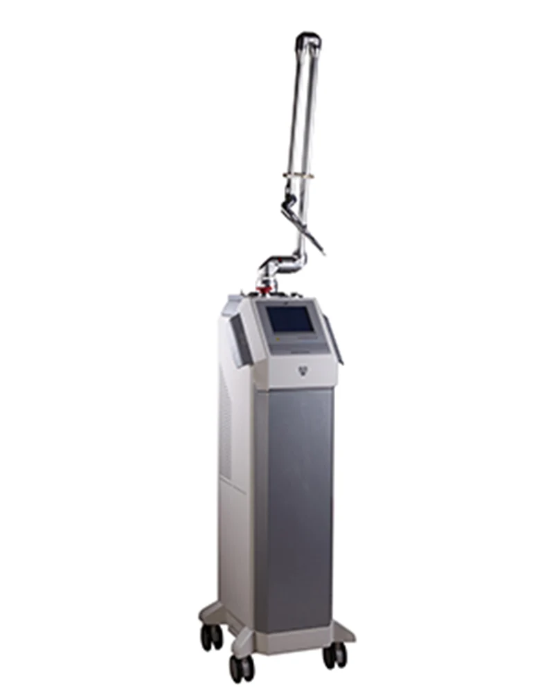
This clinic uses insulated microneedle radiofrequency for syringoma. The clinic's own treatment page describes it as delivering radiofrequency energy only at the needle tip while a specially insulated needle protects the skin surface. This page records only what has been reported about the method, not about a device brand name — clinical literature searchable under a brand name was not found.
Two domestic reports address the method itself: a retrospective review of the medical records, questionnaires, and clinical photographs of 55 patients with periorbital syringoma, and a single case in which a 30-year-old woman with lesions on the neck and axillae was compared side to side. Neither used randomization or a control group, and the second is the record of one patient with lesions that were not periorbital. Needle specifications, insertion depth, and energy settings have not been confirmed to the degree needed to print them here, and are decided at an in-person consultation.
This method has one clear absolute contraindication — carrying a pacemaker or an implanted defibrillator. A study of radiofrequency treatment describes these as 'an absolute contraindication to radiofrequency treatment' and excluded such patients. Any cardiac device in the body has to be disclosed before the procedure. Literature on the same class of devices also lists as contraindications: active infection or an uncontrolled skin condition such as eczema or psoriasis at the treatment site, a blood disorder or ongoing anticoagulant therapy, a strong tendency to keloid formation, and ongoing chemotherapy or radiotherapy. Reactivation of herpes simplex after treatment is also described, so a history of cold sores should be disclosed in advance. An expert consensus statement holds that assessment before periorbital procedures should include screening for ocular contraindications such as corneal disease or eyelid laxity — but that statement addresses laser procedures, and whether the same criteria apply to radiofrequency microneedle devices has not been confirmed. During pregnancy and breastfeeding there are no controlled studies establishing the safety of this procedure, so elective treatment is customarily deferred. In addition, no literature specifying contraindications for syringoma treatment as such was found — the items above come from literature on the same class of devices and on the safety of periorbital procedures, and the cases in which this clinic in fact does not treat are confirmed at an in-person consultation.
What to expect and how long it takes
This is not a procedure that ends in one visit. On the point that repetition is assumed, several reports agree. In a retrospective analysis of 55 patients with periorbital syringoma, about half (50.9%) showed marked improvement after a single session, and with each repeated session the lesion clearance rate rose and severity fell. That figure comes from a retrospective analysis with neither randomization nor a control group, and it is 'the proportion of patients showing marked improvement after one session', not a complete clearance rate — and that study did not assess recurrence. In a 10-patient study using a CO2 laser, 4 patients required retreatment.
There is no standard number of sessions or interval in the literature. The review of the laser literature concludes that 'there is no consensus on optimal settings', and individual reports use differing conditions such as follow-up at 6-week intervals or 3 sessions. How many sessions this clinic actually performs, and at what interval, depends on the size, number, and location of the lesions and is decided at an in-person consultation. Quantified values for when improvement begins to show and how long recovery takes were not confirmed either, so they are not printed here as figures.
What belongs alongside the course are local reactions and pigment changes. In the retrospective analysis of 55 patients above, no persistent treatment-related adverse events beyond transient erythema or crusting were reported — but it is an uncontrolled retrospective analysis, and 55 patients is not a number sufficient to detect rare events, so it is not a figure to read as establishing safety. In another small study using electrocoagulation around the eyes, burning sensation, swelling, redness, and pigment changes in the periorbital area were the most common adverse events. The complications described for destructive procedures are scarring, infection, hyperpigmentation or hypopigmentation, and recurrence. On scarring the literature is divided — small studies of 10 and 11 patients using a CO2 laser reported no scarring, while a case of hypertrophic scarring after a single CO2 laser session has been reported. On pigment change the direction is clearer: a Korean study noted that scarring, recurrence, and post-inflammatory hyperpigmentation can be particularly problematic in East Asian skin undergoing multiple treatments, and a higher Fitzpatrick skin type is treated as a concern for increased risk of hyperpigmentation and scarring with energy-based devices.
And the part that has to be stated most honestly in this condition is recurrence. Recurrence is reported regardless of the method used, and how often it occurs has not been quantified in the literature. The statement that 'recurrence is frequent' appears repeatedly in the background of many papers, but no study presenting a recurrence rate with a defined denominator and follow-up period was found; conversely, the reports stating that 'there was no recurrence' are all small case series of 12 or fewer patients with follow-up of 24 months or less. So this page states neither that it does not recur nor what percentage recurs. Following the course with the understanding that lesions can come back up in the same place even after improvement is closer to what actually happens.
What changes by area
The favored location is around the eyes, with a marked preference for the lower eyelid. In the cohort of 61 Korean patients with histologically confirmed syringoma, the most commonly involved site was the eyelid, at 43 cases (70.5%). Beyond that it also appears on the forehead, cheeks, axillae, and genital area.
The eruptive variant, which spreads widely over the trunk, is distributed differently. In a study summarizing 90 cases diagnosed at one hospital over 12 years, the mean age at diagnosis was 28.8 years, and the three most common sites were the neck, chest, and abdomen. Those figures describe 90 cases of the eruptive variant and are not values to transfer directly to the common form confined to the periorbital area.
How the intensity or the approach of treatment changes by area has not been confirmed to the degree needed to print it here. It is decided at an in-person consultation.
Other conditions that look similar
Nor is a small bump around the eye necessarily one of the two — syringoma or milia. Among the entities standard references list in the differential diagnosis of syringoma, those that genuinely overlap in this location include flat warts, xanthelasma, sebaceous hyperplasia, and angiofibroma.
All of these overlap in appearance, so telling them apart on your own by color, size, and number is difficult. What has to be done differs depending on which one it is, so the starting point is always identification.
Frequently asked questions
Does syringoma go away on its own?
The common form — multiple facial lesions — is described as tending to persist unless treated. In the eruptive variant that spreads widely over the trunk, the possibility of spontaneous regression over several years has been reported, but that is not something that transfers directly to the common periorbital form.
How is it different from milia?
Milia are keratin trapped in a sac, so there are contents to be emptied out. Syringoma is proliferated tissue derived from sweat ducts, so the tissue itself has to be removed, and whatever is left comes back up. That said, a milium-like variant of syringoma has been reported, so telling them apart by appearance alone is difficult. Identification is made at examination.
Does one session clear it?
Repetition is assumed. In a retrospective analysis of 55 patients with periorbital lesions, about half showed marked improvement after one session, and the lesion clearance rate rose with each repetition. In a 10-patient study using a CO2 laser, 4 patients underwent retreatment. There is no standard number of sessions or interval in the literature.
Will it come back?
It can. Multiple papers describe recurrence of syringoma as frequent, and standard references state that because the lesion lies within the dermis it can recur after superficial treatment. At the same time, there is no recurrence figure in the literature with a defined denominator and follow-up period. So this page states neither that it does not recur nor what percentage recurs.
It is right next to the eye — is the eye safe?
In periorbital procedures, protecting the eye is part of the procedure. The fact that the most common site of syringoma is the lower eyelid changes the risks of this treatment directly. A consensus statement from a panel of dermatologic laser surgeons and ophthalmologists states that eye shields are mandatory for all laser procedures performed within the bony orbit, because every laser wavelength poses some degree of risk to ocular structures. The reported risks include corneal burns, ectropion, infection, dry eye, lamellar keratitis, cataract, temporary or permanent vision loss, and corneal and scleral scarring. The eyelid is described as not thick enough to protect the eye from injury, so a metal shield must be worn throughout the procedure, and the procedure is stopped immediately if ocular pain or a change in vision occurs during it. These criteria were, however, established for lasers, and literature on ocular safety standards for radiofrequency microneedle devices was not found. How this clinic provides protection is explained at the consultation before treatment.
Could it be cancer?
No case of a lesion confirmed as syringoma undergoing malignant change has been reported. There are, however, malignant adnexal tumors that are confused with syringoma both clinically and histologically, and cases of malignant tumors on the eyelid that appeared benign have been reported. Among the three eyelid cases reported, one patient required five excisions to achieve complete removal, and another recurred and was advised to undergo more extensive surgery. This is rare. But a procedure that destroys a lesion cannot be undone, so establishing what it is by examination comes first.
This page is general information about a treatment and is not a diagnosis or a recommendation. The right choice differs by skin condition, so treatment is decided after an in-person examination. Individual results vary.
References
- Syringoma StatPearls, last updated 2024-03-20 (Geist R, Crane JS)
- Syringoma DermNet, last reviewed 2024-12 (Singh A, Oakley A)
- Evaluation and management of the patient with multiple syringomas: A systematic review of the literature J Am Acad Dermatol 2016;74(6):1234-1240.e9 (Williams K, Shinkai K)
- Syringoma: a clinicopathologic and immunohistologic study and results of treatment Yonsei Med J 2007;48(1):35-40 (Lee JH, Chang JY, Lee KH)
- Eruptive syringomas: Summary of ninety cases and a brief literature review J Cosmet Dermatol 2023;22(3):1128-1133 (Lei H, Wang Z, Ma X, Zhang Z, Feng Y, Zheng Y)
- Milium, milia DermNet, published 2009 (Gupta M, Oakley A)
- Milium-like syringoma: a case study on histogenesis J Cutan Pathol 2004;31(4):336-40 (Wang KH, Chu JS, Lin YH, Hu CH, Lee WR)
- Microcystic Adnexal Carcinoma StatPearls, last updated 2023-04-24 (Zito PM, Mazzoni T)
- Microcystic Adnexal Carcinoma: A Review of the Literature Dermatol Surg 2017;43(8):1012-1016 (Gordon S, Fischer C, Martin A, Rosman IS, Council ML)
- Syringomatous carcinoma of the eyelid and orbit: a clinical and histopathological challenge Br J Ophthalmol 1997;81(8):668-72 (Hoppenreijs VP, Reuser TT, Mooy CM, de Keizer RJ, Mourits MP)
- Efficacy and safety of lasers in treating syringomas: a review of the literature J Dermatolog Treat 2022;33(8):3127-3135 (Alsaidan MS)
- A new treatment for syringoma. Combination of carbon dioxide laser and trichloroacetic acid Dermatol Surg 1998;24(12):1370-4 (Kang WH, Kim NS, Kim YB, Shim WC)
- The treatment of syringomas by CO2 laser using a multiple-drilling method Dermatol Surg 2007;33(3):310-3 (Park HJ, Lee DY, Lee JH, Yang JM, Lee ES, Kim WS)
- Intralesional electrodesiccation of syringomas Dermatol Surg 1997;23(10):921-4 (Karam P, Benedetto AV)
- Intralesional Electrocoagulation With Insulated Microneedle for the Treatment of Periorbital Syringomas: A Retrospective Analysis Aesthet Surg J 2021;41(4):490-498 (Ahn GR, Jeong GJ, Kim JM, Hong JY, Li K, Lee CK, Kim BJ)
- Comparison of microinsulated needle radiofrequency and carbon dioxide laser ablation for the treatment of syringoma Dermatol Ther 2019;32(3):e12912 (Bae JY, Jang DH, Lee JI, Jung HJ, Ahn JY, Park MY)
- Expert Consensus on Ocular Safety During Laser Procedures: A Practical Guide to Eye Safety by a Panel of Dermatologist Laser Surgeons and Ophthalmologists J Cosmet Dermatol 2025;24(7):e70339 (Kesty K, Kesty C, Benson J)
- Ocular Injury in Cosmetic Laser Treatments of the Face J Clin Aesthet Dermatol 2018;11(2):15-18 (Huang A, Phillips A, Adar T, Hui A)
- Cosmetic Treatments with Energy-Based Devices in Skin of Color Facial Plast Surg 2023;39(5):496-500 (Chao JR, Porter JP, Hessler J)
- Treatment of multiple facial syringomas with the carbon dioxide (CO2) laser Dermatol Surg 1999;25(2):136-9 (Wang JI, Roenigk HH Jr)
- Periorbital syringoma: a pilot study of the efficacy of low-voltage electrocoagulation Dermatol Surg 2006;32(10):1244-50 (Al Aradi IK)
- Microneedling: Advances and widening horizons Indian Dermatol Online J 2016;7(4):244-54 (Singh A, Yadav S)
- Treatment of facial skin laxity by a new monopolar radiofrequency device J Cutan Aesthet Surg 2011;4(1):7-11 (Wollina U)
- A review of the safety of cosmetic procedures during pregnancy and lactation Int J Womens Dermatol 2017;3(1):6-10 (Trivedi MK, Kroumpouzos G, Murase JE)
Related information
- What are milia? — What milia are and how they are dealt with is covered in that article
- Syringoma & Milia clinic — This clinic's syringoma and milia care
- Under Eye Area treatments — The area below the eye, where syringoma most commonly appears