
Pigmentation • Differential diagnosis
Lentigo: What It Is and How It Differs from Flat Warts
- Written by
- The Other Dermatology medical team
- Published
- 2026.08.21
When brown spots start turning up on the face one after another, most people think of age spots first. In the exam room, though, what patients call an age spot turns out to be several different things. It may be pigment laid down by ultraviolet light, it may be a viral infection — flat warts — and less often it may be an early skin cancer. What differs is not how it is removed, but **whether it should be removed at all.** Read the lesion wrong and removing it takes the result in the opposite direction.
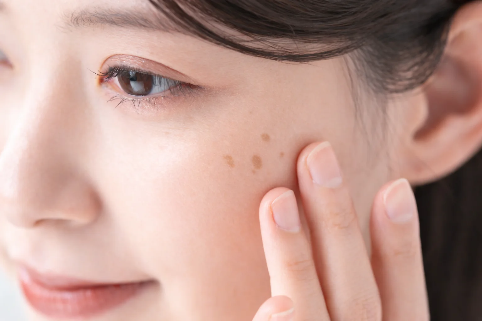
What is Lentigo?
"Age spot" is not a diagnosis. It is an everyday word for the brown patches that appear on the face, the backs of the hands and the arms, and in the exam room the word usually points to one of two lesions — solar lentigo and seborrhoeic keratosis. Usually is the operative word, and that is the point of this article. Other things present as the same brown patch and are neither of these two, and early skin cancer is among them. We come back to it under "Other conditions that look similar" below.
Solar lentigo is a common benign pigmented macule produced by chronic ultraviolet exposure. Lesions range from a few millimetres to a few centimetres, the colour runs from light tan through dark brown to black, and the border is well defined. Histologically the change is confined to the epidermis: melanocytes producing pigment briskly are gathered in one place, with some pigment-laden cells seen in the dermis. It is common from middle age onward and uncommon under 12 years of age. In white skin it has been reported in up to 90% of people over 60, and it is common in Asian skin as well, where it is described as tending to appear on the face.
Seborrhoeic keratosis is a common sign of skin ageing that shows up in adulthood. The epidermis thickens into a sharply demarcated plaque that feels as though something had been stuck onto the skin, and the surface may be smooth, waxy, or rough like a wart. It usually begins to appear in the 30s and 40s, and more than 90% of people over 60 have at least one.
The two are less separate diseases than a continuum. Progression of a solar lentigo into a pigmented seborrhoeic keratosis over time is described in the literature, which is why it is common to find flat lesions and raised ones side by side on the same face.
This matters only once it is clear what the lesion is, but the two do share something. They are made by ultraviolet light and by time, not caught from anyone. That is where they part company with flat warts.
How is it different from Flat Warts?
Flat warts (verruca plana) are an infection with human papillomavirus (HPV), most often types 3 and 10. Flat, sharply bordered papules 2–5 mm across appear in groups on the face, the backs of the hands and the shins. They may be skin-coloured, and they may also take on a light brown tint, so a face carrying several brown macules is not easy to tell apart from age spots. Flat pigmented lesions of the face are described in the literature as an area where findings overlap even under magnification, which makes diagnosis there difficult.
The trouble comes after removal. Warts spread locally after trauma as minor as scratching. The virus seeds itself onto another site of the same skin — autoinoculation, called pseudo-Koebnerization in the literature. Cases have been reported in which existing lesions spread in a line along the micro-trauma and the broken epidermal barrier left by cosmetic procedures, and a literature review documents warts appearing anew at laser-treated sites (these are case reports, so how often this happens cannot be stated). Spread of flat warts by shaving over the beard area and the shins is a well-recognised route.
One more thing. Treating warts does not kill the virus; it removes the skin the virus is in. HPV has been detected histologically as far as 15 mm from a visible lesion, so clearing only what can be seen can leave virus in the surrounding skin. That is why removing flat warts in the frame of mind you would bring to an age spot can end with more lesions, arranged along the treatment lines, rather than fewer. Before reading the table below, one caveat — it shows the direction a differential runs; it is not a table for deciding which side your own lesion falls on. A lesion that does not sit cleanly in either column, or one that has been changing lately, belongs outside this table.
Flat Warts
- Cause
- Viral infection, HPV types 3 and 10 among others
- Number and arrangement
- 2–5 mm lesions in crops; linear arrangement along scratch marks and shaving lines is not unusual
- On palpation
- Flat, smooth papules
- After removal
- May recur from virus left behind, or spread along the treatment lines
Lentigo
- Cause
- Chronic ultraviolet exposure and skin ageing
- Number and arrangement
- A few millimetres to a few centimetres; built by accumulated ultraviolet exposure, so it does not multiply abruptly over a short period
- On palpation
- Solar lentigo is flat; seborrhoeic keratosis feels raised and slightly rough, as if stuck on
- After removal
- May return in the same place if ultraviolet exposure continues
How is it treated?
The first step of treatment is not removal. It is deciding what the lesion is.
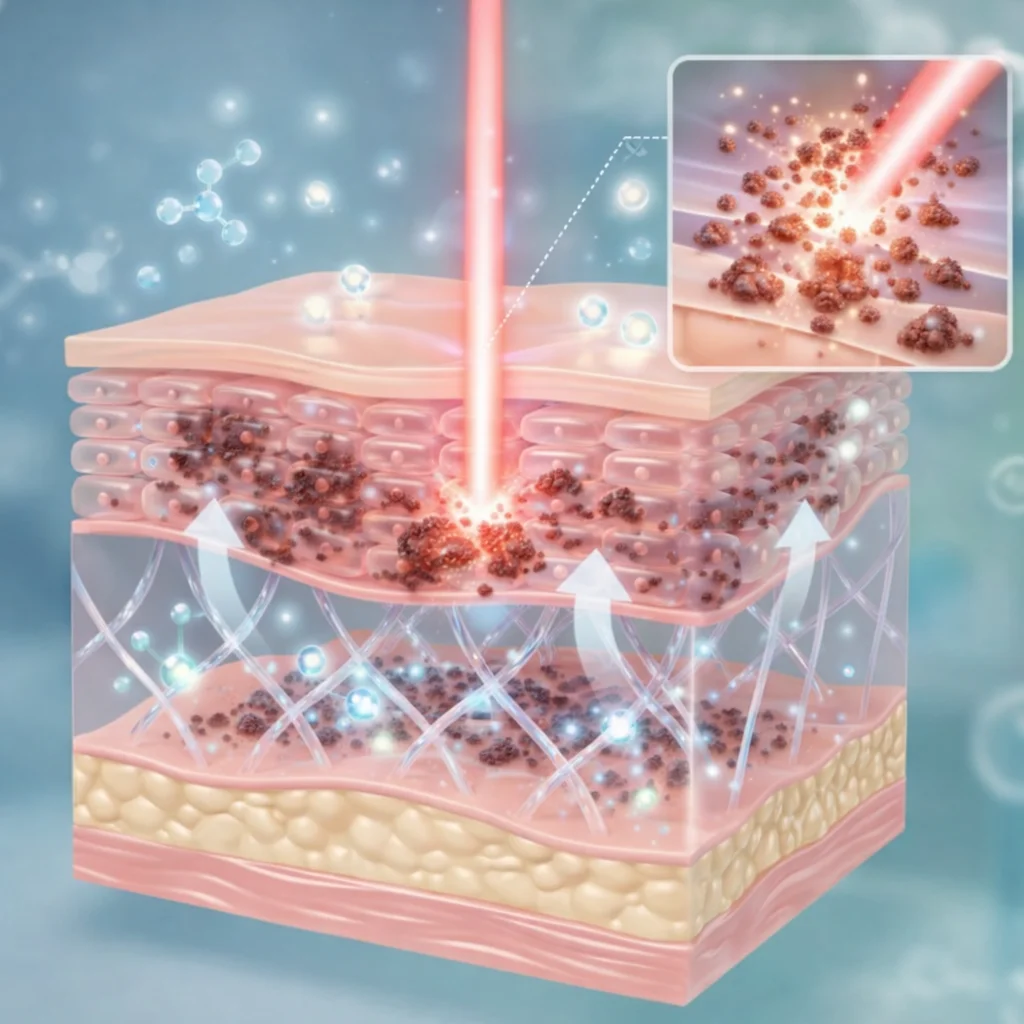
The distribution of pigment, the border and the surface structure of the lesion are assessed on examination. Flat pigmented lesions of the face are a site where clinical findings overlap one another, so if the assessment is not settled there, or if malignancy is suspected, we do not move on to removal. The literature states that a biopsy should be taken when the diagnosis is uncertain or malignancy is suspected, and adds that when the colour within a single lesion divides into several shades, separate samples should be taken from each area — different pathology can sit together in one place.
Once the lesion is confirmed to be an age spot, the plan moves toward removing a lesion confined to the epidermis. A flat solar lentigo, whose pigment sits in the epidermis, and a seborrhoeic keratosis, raised by a thickened epidermis, are the same brown to the eye but differ in what is happening in the tissue, so the approach diverges. Either way, keeping ultraviolet light off the treated site while new epidermis covers it is part of the plan. The literature names strict sun protection as the condition for preventing recurrence after treatment.
If the lesion turns out to be flat warts, the axis of the plan changes. The goal shifts from "clearing what is visible" to "reducing them without letting them spread." The literature states that no treatment is universally effective at eradicating the virus, and these are lesions that can also resolve on their own, so what to treat and how far to go becomes a matter of judgement.
The two are handled as separate items of care from the outset. Mole and age-spot removal, and flat wart and milia removal, are managed as different services.
What to expect and how long it takes
A site where a lesion has been removed goes through healing. If ultraviolet protection falls short during this period, it can lead to pigmentation. How recovery runs depends on the size, depth and site of the lesion and on skin type, so the actual schedule is set at an in-person consultation.
Removal is a wound, and a wound can leave a mark of its own. Destructive treatment carries the risk of leaving the treated site paler or darker than the surrounding skin, and that change may be temporary or it may last. Post-inflammatory hyperpigmentation is set out in the literature as a characteristic adverse effect of the CO2 laser, which delivers more thermal energy into tissue than other resurfacing lasers do. The risk of dyspigmentation and of scarring is higher in darker skin, and that is the reason laser treatment in skin of colour has historically been approached with hesitancy. In a study of persistent viral warts treated with CO2 laser, post-operative pain, temporary loss of function of the treated part, and scarring were commonly observed side effects.
Flat warts follow a different course. They are known to disappear on their own once cell-mediated immunity engages — in a series of 25 cases, every lesion underwent sudden inflammation and then regressed completely within 2–6 weeks. But there is no predicting when this will happen, or in whom. In children, warts in general are reported to clear without treatment in half of cases within 6 months and in 90% within 2 years, but those figures are for children and do not carry over to adults. In adults they persist longer.
Aftercare
The scab over a treated site is better left until it comes away by itself. Lifting it early exposes skin that has not finished healing, and the route from inflammation during healing to pigmentation is set out in the literature. How long to keep the dressing on varies with the depth and site of the lesion, so follow what you are told in the clinic.
Sun protection here is not care for the recovery period; it is the condition for preventing recurrence. Age spots can return in the same place if ultraviolet exposure continues, and ultraviolet light during healing is a direct cause of post-inflammatory hyperpigmentation. Topical steroids have been reported to lower the risk of post-inflammatory hyperpigmentation after laser treatment in Asian skin, but whether to use a medication and for how long is decided against the state of the skin.
If the lesion has been judged to be flat warts, one more item joins the list. Touching and scratching are themselves the route by which they spread. Do not rub the area or pick at the flaking skin, and if lesions sit where you shave — the beard area or the shins — how and when to shave is worked out together at the consultation. Not sharing towels, razors and similar items with other people belongs to the same reasoning.
What changes by area
The face is where the two lesions overlap most often. Solar lentigo is common in Asian skin too and tends to appear on the face, and the face is a classic site for flat warts as well. Lentigo maligna is likewise described as presenting on chronically sun-damaged sites such as the head and neck, as a flat hyperpigmented macule — the face is where all three converge.
The backs of the hands, the forearms and the shins are where the paths separate. The backs of the hands, the forearms and the upper chest take accumulated ultraviolet exposure directly and are common sites for solar lentigo, while the shins and the beard area of the face are named in the literature as the sites where shaving spreads flat warts. Flat brown papules increasing in a line across an area you shave is not a change that accumulated ultraviolet exposure explains.
Other conditions that look similar
Early skin cancer comes first on this list. Lentigo maligna is described in the literature as overlapping in appearance with macular seborrhoeic keratosis, pigmented actinic keratosis and solar lentigo, and as difficult to diagnose even for experienced dermatologists. Seborrhoeic keratosis in turn can resemble basal cell carcinoma, squamous cell carcinoma and melanoma, particularly among pigmented lesions on exposed sites such as the face. Unlike the two entries below, here the delay in telling them apart is itself the harm, so where the assessment is ambiguous, confirming what the lesion is comes before removing it. If it is not certain what it is, it is not removed. In a retrospective analysis of 503 patients with biopsy-confirmed lentigo maligna, 37 (7.4%) had received cosmetic treatment before the diagnosis, and 6 of those were later found to have invasive cancer. A lesion whose size, shape or colour has been changing lately, one with an irregular border, one whose colour divides into several shades within the same lesion, or one that repeatedly forms a crust that will not heal, needs confirmation before it needs a laser.
Freckles (ephelides) are also frequently confused with age spots. Both are brown macules, but their origins differ. Freckles arise on a genetically determined background with sunlight pulling the trigger; solar lentigo is made by accumulated ultraviolet exposure and photoageing themselves. The differences the literature cites are age and season. Freckles first appear in childhood, increase into young adulthood, and fade in winter, then darken again in summer. Solar lentigo does not fade in winter; it persists through the year and darkens only slightly in summer.
Milia are confused by texture rather than colour. They are keratin-filled cysts under 3 mm sitting beneath the epidermis, appearing as white or pale yellow dome-shaped papules on the cheeks, eyelids and forehead. Like flat warts, they scatter across the face in numbers, so by count alone they can look similar — but they are neither a pigmented lesion nor a viral infection. They are divided into primary milia, which arise spontaneously, and secondary milia, which follow trauma, medication or blistering disease.
Frequently asked questions
My age spots increased suddenly — why can I not just have them removed?
If the count is rising quickly, "age spots built by accumulated ultraviolet exposure" does not account for it on its own. Flat warts spreading by autoinoculation are the classic example, but not the only one, so the sudden increase is itself a sign to have checked at a consultation. What the lesions are is decided by looking at them directly.
Do flat warts ever go away on their own?
That course is described in the literature. In a series of 25 cases, every lesion underwent sudden inflammation and then regressed completely within 2–6 weeks, which is explained as a cell-mediated immune phenomenon. But there is no predicting when it will happen or in whom, so it is hard to build a plan around waiting for it. In children, clearance of warts in general within 2 years is reported at 90%, but that figure is for children and does not carry over to adults.
Do I really need a biopsy?
Not for every lesion. But where the assessment is ambiguous — a lesion that has been changing lately, one whose colour divides into several shades, one with an irregular border or with bleeding or ulceration — confirming what it is before removing it is the order the literature recommends. If a laser clears it first, there is no tissue left to examine.
Are flat warts contagious to other people?
They are an HPV infection, so they can be passed on. What is more common in practice, though, is autoinoculation onto another site of your own skin. Not sharing towels and razors, and not rubbing the area or picking at flaking skin, are the practical ways to reduce spread.
Will they come back after removal?
Age spots can reappear in the same place or nearby if ultraviolet exposure continues. The literature names strict sun protection as the condition for preventing recurrence after treatment. Flat warts recur differently again, because virus may remain in surrounding skin that is not visible.
How do I tell freckles from age spots?
The differences the literature cites are age and season. Freckles appear from childhood and increase into young adulthood, fading in winter and darkening again in summer. Age spots (solar lentigines) appear from middle age onward and hold through the year without fading in winter. This distinction only means something once it is clear what the lesion is, though — a lesion that has been changing lately needs checking at a consultation regardless of the season.
This page is general information about a treatment and is not a diagnosis or a recommendation. The right choice differs by skin condition, so treatment is decided after an in-person examination. Individual results vary.
References
- Solar lentigo DermNet NZ
- Seborrhoeic keratosis DermNet NZ
- Melanoma — ABCDE warning signs DermNet NZ
- Lentigo maligna part I: Epidemiology, risk factors, and diagnosis J Am Acad Dermatol 2024
- Lentigo maligna — analysis of cases treated cosmetically before diagnosis Lasers Surg Med 2017
- Cosmetic warts: pseudo-Koebnerization of warts after cosmetic procedures J Clin Aesthet Dermatol 2015;8(7)
- Koebner Phenomenon StatPearls
- Post-inflammatory hyperpigmentation after carbon dioxide laser: review of prevention and risk factors PubMed
- Outcome of carbon dioxide laser therapy for persistent cutaneous viral warts PubMed
- Spontaneous regression of plane warts after inflammation: clinical and histologic studies in 25 cases PubMed
- Sun-induced freckling: ephelides and solar lentigines Pigment Cell Melanoma Res 2014
- Milia StatPearls
- Viral wart DermNet NZ
Related information
Related conditions
- What melasma is, and how it differs from ABNOM — Pigment that can be mixed in with lentigines
Treatment pages
- Lentigo and pigmented lesion treatment — How dark, deep-seated lentigines are approached
- Syringoma and milia — telling them apart and treating them — When multiple facial papules are distinguished by texture
- Melasma treatment and management — A pigmentary condition where management, not removal, is the axis